Your Visit

Preserve Your Vision with Comprehensive Eye Exams

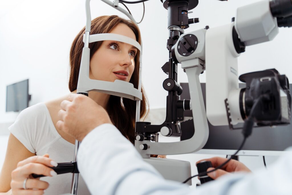
Your eyes deserve the best care. A thorough eye examination is essential for maintaining optimal eye health and catching potential issues early. 
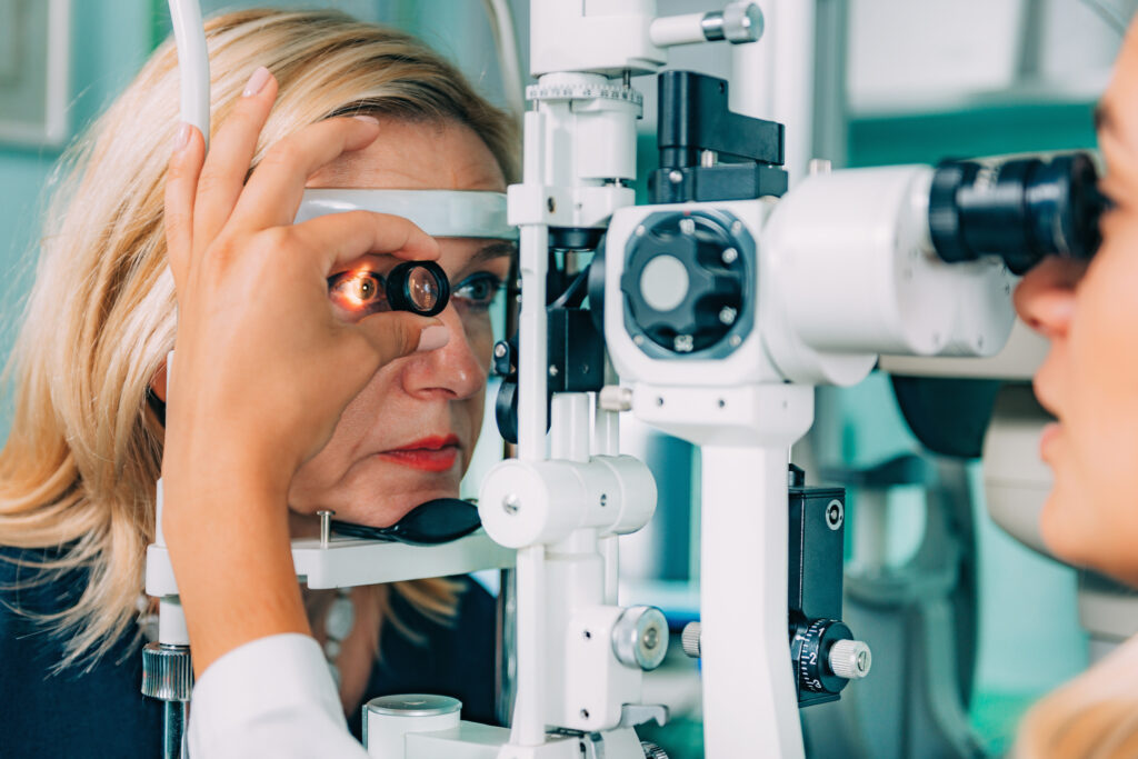
Our experienced ophthalmologist will evaluate your overall eye health and vision, ensuring you receive the highest quality care. Here’s what to expect during your visit and the advanced diagnostic tools we use:

What to Expect During Your Eye Exam

  1. Visual Acuity Test: Measures how clearly you see at various distances using an eye chart.
  2. Slit-Lamp Examination: Provides a detailed view of the front structures of your eye, including the cornea, iris, and lens.
  3. Tonometry: Measures the pressure inside your eyes to screen for glaucoma.
  4. Pupil Dilation: Enlarges your pupils for a comprehensive examination of the retina and optic nerve.

Advanced Diagnostic Tools for Eye Conditions

Glaucoma Detection

  1. Optical Coherence Tomography (OCT): Non-invasive imaging that captures cross-section pictures of your retina, detecting and monitoring glaucoma by measuring retinal nerve fiber thickness.
  2. Heidelberg Retinal Tomography (HRT): Provides detailed 3D images of the optic nerve head, identifying early glaucomatous damage.
  3. Visual Field Test: Maps your field of vision to detect blind spots and peripheral vision loss, common signs of glaucoma.

Cataract Evaluation

  1. Slit-Lamp Examination: Reveals the formation and extent of cataracts.
  2. Retinal Examination: After dilation, examines the back of the eye for cataract severity and other potential issues.
  3. Biometry (A-Scan and B-Scan Ultrasound): Measures the eye’s length and curvature, essential for selecting the right intraocular lens for cataract surgery.

Dry Eye Diagnosis

  1. Tear Film Break-Up Time (TBUT): Measures the stability of the tear film to diagnose dry eye syndrome.
  2. Schirmer Test: Assesses tear production to determine if your eyes produce enough moisture.
  3. Meibomian Gland Evaluation: Evaluates the glands that produce the oily layer of the tear film, often linked to dry eye syndrome.

Additional Advanced Diagnostic Tools

  1. Corneal Topography: Maps the cornea’s surface curvature to detect vision-affecting irregularities.
  2. Fundus Photography: Captures detailed images of the retina for diagnosing and monitoring various retinal conditions.
  3. Pachymetry: Measures corneal thickness, crucial for glaucoma management.
  4. Anterior Segment OCT: Provides detailed images of the front part of the eye, aiding in diagnosing conditions like angle-closure glaucoma.

Experience the Latest in Eye Care Technology

At our practice, we are committed to using the latest technology to ensure precise diagnosis and effective treatment. Regular eye exams are vital for preserving your vision and eye health. Schedule an appointment today to experience comprehensive care tailored to your needs. Don’t wait—take the first step towards maintaining your eye health and clear vision.